249 – Carcinoid tumor
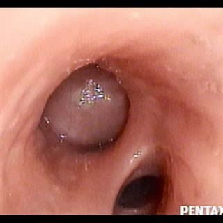
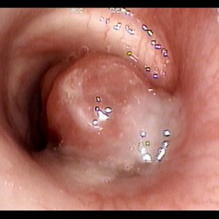
Carcinoid tumor that appears with a fairly classic appearance. Due to its smooth convexity and its free contact with the bronchial walls it is compared with a “glove finger”.

Carcinoid tumor that appears with a fairly classic appearance. Due to its smooth convexity and its free contact with the bronchial walls it is compared with a “glove finger”.
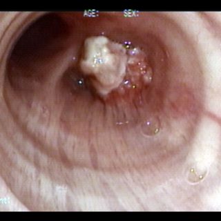
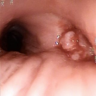
Blend of coloration: exophytic lesion showing an irregular and pinkish polylobulated area while its most prominent end is creamy white, as a product of necrosis in a distal area of the tumor.
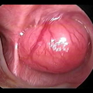
“Threatening” aspect. With its reddish coloration that declares a sufficient vascularization, and its thick vessels that crown the visible hemisphere of this tumor that from the entrance of the right source bronchus, it challenges the bronchoscopist, tempting him to defer the biopsy to a safer time. A rigid bronchoscopy will make it possible to thermocoagulate … Read more
After implanting this “Y” shaped stent, the advance of tumor growth into the stent makes it emerge through its right bronchial branch. As an undesired consequence of the subocclusion of this source bronchus, there is an accumulation of secretions at its entrance, indicating a precarious airflow and an ineffective cough.
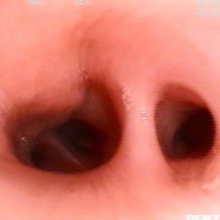
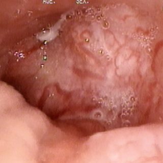
Example of “intramural” injury. The tumor deforms the light by occupying the wall without destroying the mucosa yet. Positive biopsy. Undifferentiated carcinoma.
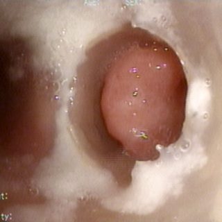
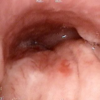
This carcinoma occurs in the trachea with a very smooth surface which will suspect a carcinoid tumor. Note the intense mucosal vascularization that with a more or less parallel disposition is directed to the tumor.
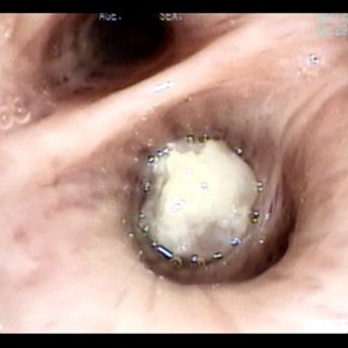
Another form of presentation of the endobronchial tumor that occurs frequently: only whitish formation. It is no more than necrotic tissue on the surface because it is the furthest point from its base of vascularized implantation. The secretions cover it and can dry out due to the disturbance in the airflow produced by the tumor.
Vegetative tumoral formation that occludes the right upper lobe, which also shows a noticeable widening of its spur. The lesion appears with freshly emitted blood due solely to the cough that occurred during the endoscopic exploration. Insufficient local anesthesia will trigger coughing during the examination and spontaneous bleeding will make it difficult to see and … Read more
Close-up image of a bulky tumor that completely obstructs the bronchial lumen. Note the particular arrangement of the surface vasculature. Some vessels interrupt their journey intermittently. It is another expression of the “vascular stop” because of the tumoral infiltration of its wall.
Locoregional neoplastic disease. Although there is no lesion of endoluminal growth, the bronchial mucosa is decidedly affected by irregular edema and visible thickening. The rigidity and decreased mobility are other signs that can be seen during endoscopy when the bronchus is “fixed” to the tumor that surrounds it.