
larynx

009 – Cordal Paralysis in Adduction
Bilateral chordal paralysis in adduction. It is the most frequent congenital paralysis and in 50% of the cases is associated with other neurological alterations The remaining 50% is idiopathic and reverses in the first year of life. (Hospital Pediatrics Juan P. Garraham).
008 – Thickening Cordal
Bilateral thoracic thickening that seems to occlude the Morgagni ventricles.

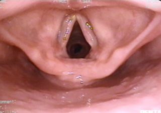
005 – The Larynx
View of the larynx. The bronchoscope has been located in the midline, raising the free edge of the epiglottis and remains supported on its pharyngeal side. The vocal cords are in abduction. The first tracheal ring can be visualized through the glottic cleft.
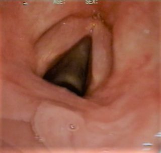
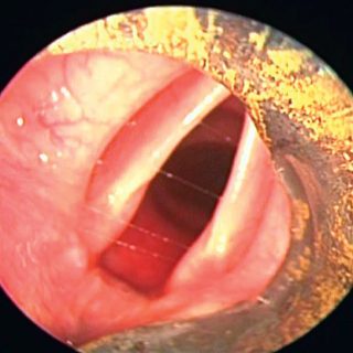
004 – Strings in Abduction
View of the normal laryngeal crown, with the vocal chords in an abduction position.
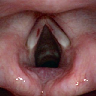
003 – The Larynx
Larynx: it can be clearly seen the arytenoepiglottic folds and vocal cords in adduction. The edge of the left vocal cord isirregular in its middle third.
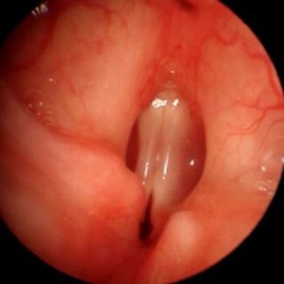
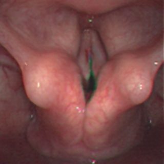
002 – Vocal Strings
The bronchoscope has overcome the epiglottis and the arytenoids, now facing the vocal cords, here in a paramedian position. Third anatomical repair for intubation. It is the right time to start the 90º rotation of the bronchoscope to separate the vocal cords with the bevel and allow smooth entry into the trachea.
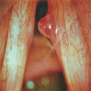
001 – Edge of Epiglotis
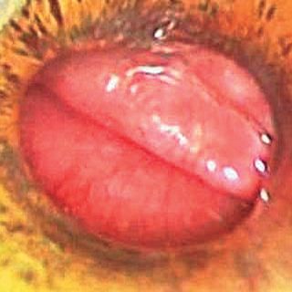
After the uvula, the free edge of the epiglottis is the second point of reference for a bronchoscopist during orotracheal intubation.