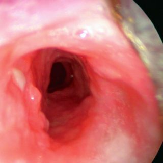
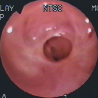
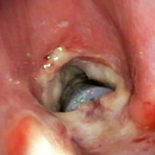
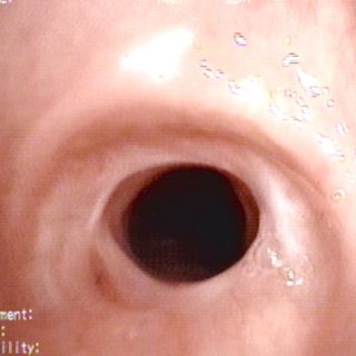
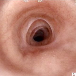
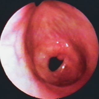
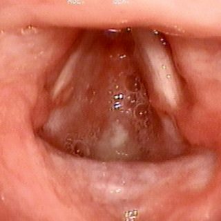
194 – Tracheal Stenosis
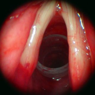
Silicone stent acting as a support in a tracheal stenosis at the level of the first rings. The vocal cords are somewhat thickened and with obvious congestion in the mucosa in its posterior third, close to the arytenoids.