206 – Spine Spreading
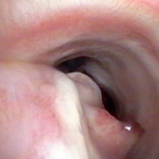
Stent implanted in intermediate bronchus. Note the uneven widening of the spur of the right upper lobe.

Stent implanted in intermediate bronchus. Note the uneven widening of the spur of the right upper lobe.
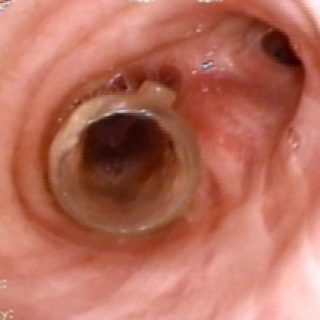
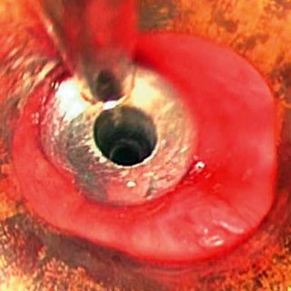
After implanting this “Y” shaped stent, the advance of tumor growth into the stent makes it emerge through its right bronchial branch. As an undesired consequence of the subocclusion of this source bronchus, there is an accumulation of secretions at its entrance, indicating a precarious airflow and an ineffective cough.
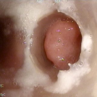
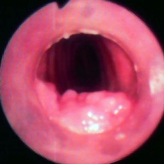
Central and critical tracheal stenosis. Smooth and tense appearance due to congestive edema of the mucosa. Cetrángolo Hospital
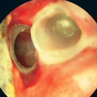
Double stent: after the endo-surgical resection of an extensive carcinoma, two silicone stent were applied in the tracheal bifurcation, leaving the carina “enclosed” between them. In the image, the generalized edema dominates the field and very abundant mucopurulent secretions accumulate around the prosthesis and inside one of them, in which a bubble has formed at … Read more
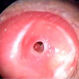
Appearance of the tracheal mucosa after the removal of a stent. There is an arch of residual stenosis, still congestive, and a mucosal bed with bulging edema due to prolonged contact with the prosthesis in that area.
The metallic olive crosses the narrowness of the stenosis gently, forcing the opening of the tracheal lumen. The central hole of the instrument allows the passage of air preventing the complete interruption of ventilation during the procedure.
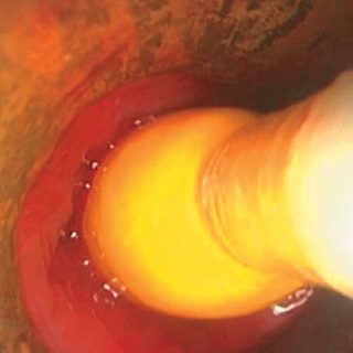
A dilation balloon full of saline solution, exerts circumferential pressure during dilation of a stenosis. The appearance of the tracheal wall, edematous and congestive, can be appreciated after decompression of the elastic balloon. Note the distance between the balloon and the mucosa as a result of the progressive dilation maneuver.
These smooth protrusions that seem to pile on the posterior tracheal wall or mucous membrane, correspond to granulomas originated by a fearsome situation: the “tracheal cannula tip injury”. The lack of stable fixation of the pavilion of the tracheostomy cannula against the neck, allows movements of the device that tends to swing on its support in … Read more
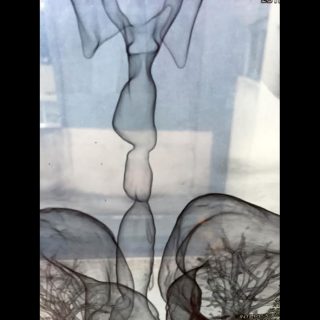
A rare form of complex stenosis is double stenosis. Their treatment differs considerably according to their anatomical location: very separate, close together or even worse, as seen in the reconstruction image: “neither together nor separated”.
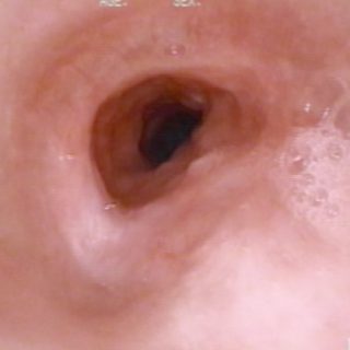
Tracheal Stent after 10 months remain implanted. The walls of the endoprosthesis are free of secretions and incrustations, but several granulomas have developed in the mucosa close to its distal end. One, very bulky and bulgy, sits on the entire back wall.