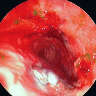
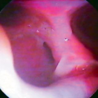
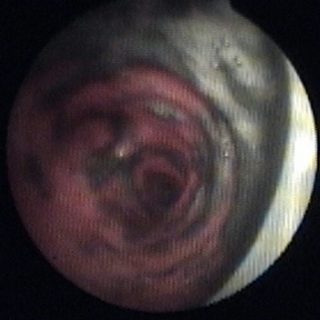
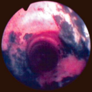
243 – Myofibroblastic Bronchial Tumor

243 - Myofibroblastic or pseudoinflammatory tumor

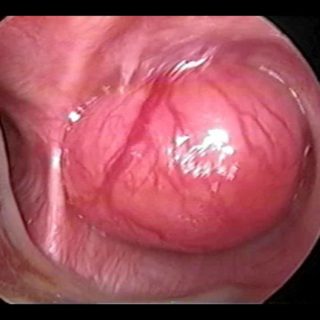
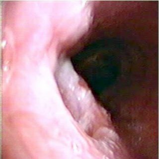
“Threatening” aspect. With its reddish coloration that declares a sufficient vascularization, and its thick vessels that crown the visible hemisphere of this tumor that from the entrance of the right source bronchus, it challenges the bronchoscopist, tempting him to defer the biopsy to a safer time. A rigid bronchoscopy will make it possible to thermocoagulate … Read more

242 – Tracheal Fistula

242 - Tracheal fistula

Here the communication with the esophagus is so wide that it occupies the entire diameter of the tracheal posterior wall. The condition responds to a long list of causes that include therapeutic accidents and is favored by the chronic prostration and poor nutrition of these patients. Cetrángolo Hospital

238 – Tracheal Fissure

238 - Tracheoesophageal fistula

Tracheoesophageal fistula: Intense edema of the entire tracheal mucosa, with extensive areas of submucosal hemorrhage. The edema becomes irregular in the posterior wall and a U-shaped fissure occupies the center of the image. The dark coloration of its path is due to the methylene blue instilled in the esophagus for its detection. Isidoro R., Sinus … Read more

235 – Airway burn

235 - Airway burn

The dark and patchy coloration that becomes confluent in all the trachea closest to this image, is due to the presence of abundant accumulated carbon particles and combined with mucus. They were inhaled during a fire and may exist with or without airway burn. The endoscopic examination is “poorly lit”; this effect is a consequence … Read more

233 – Tracheostomy. Ostoma Tutor

233 - Ostoma Tutor

Improper position of a Tutor. Its inner end has separated from the edge of the ostoma and occupies part of the tracheal lumen. It must be corrected by pulling it from its outer branch and then fixing it in the proper position with its retaining ring.