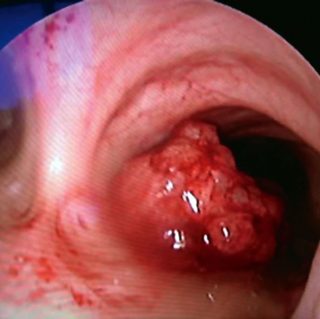
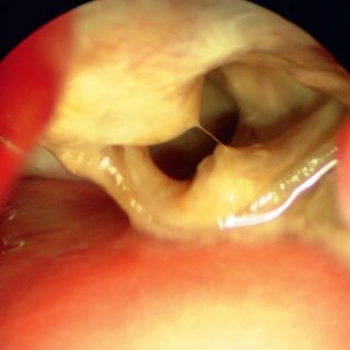
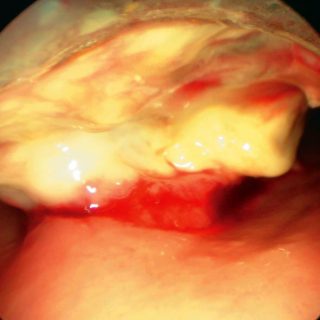
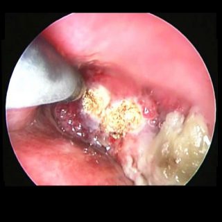
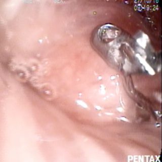
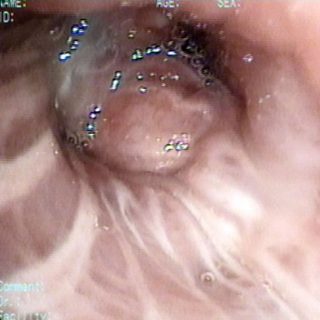
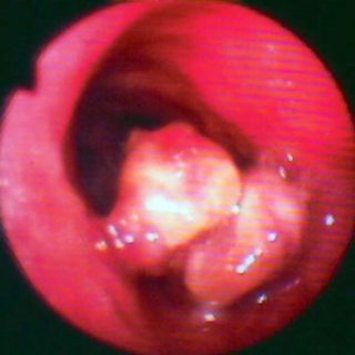
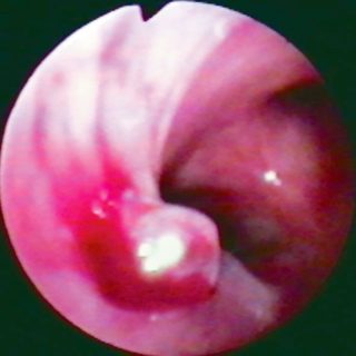
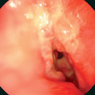
274 – Tracheal Carcinoma

274 - Giant cell carcinoma I

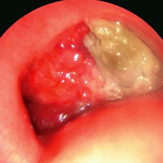
A voluminous vegetative formation settles in the tracheal carina and completely occludes the light of the right source bronchus and reduces that of the left. The surface is irregular, with protrusions and increased vascularization in some areas. The tissue that obstructs the right main bronchus has been necrotic. The posterior tracheal wall seems to be … Read more

272 – Tracheal Carcinoma

272 - Giant cell carcinoma

Trachea: a giant cell carcinoma has completely infiltrated the wall. Two ends of the cartilaginous rings are visible in the light as a result of the destruction of their mucosa, now covered by thick purulent secretions and suffering continuous dehydration due to being exposed to the turbulent airflow caused by local narrowness and inflammation.

269 – Bronchial Tumor

269 - Endobronchial tumor

Although the foreground magnifies the image, it is only the small biopsy forceps with fenestrae in its leaflets, commonly used with the flexible bronchoscope, doing its work of tissue capture for the diagnosis of this endobronchial tumor.

267 – Tracheal Tumor

267 - Tracheal Tumor

Tracheal tumor with double origin: two large formations occupy the center of the tracheal lumen, although there is still a sufficient area for ventilation. In the image, the formations appear superimposed, but they have an independent origin constituted by pedicles that emerge from the tracheal wall. Both correspond to a single primary carcinoma.

266 – Bronchial Carcinoma

266 - Bronchial Carcinoma

Small “bronchial bulge” very erect. Remember better a rock. It is located at the end of the left source bronchus, near the crossroads and results from the invasion of the bronchial wall by a carcinoma. The mucosa is pulled, forming grooves. Further, the widened spur suggests underlying adenomegalies.

265 – Intramural Bronchial Tumor

265 - Carcinoma

Bronchial light “stabbing”: an “intramural, intraluminal and infiltrating” carcinoma deforms the light from the left source bronchus. The intense edema and thickening of the bronchial mucosa contribute to the distortion and the marked reduction of the caliber. On the left wall it acquires a cordoned appearance that forms mamelons as it ascends and occupies the … Read more