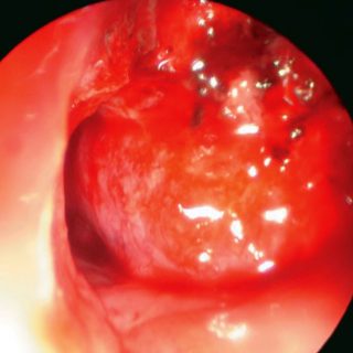
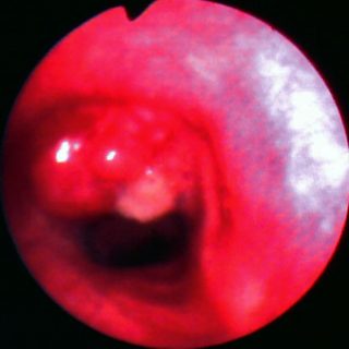
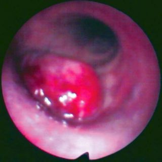
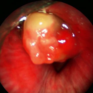
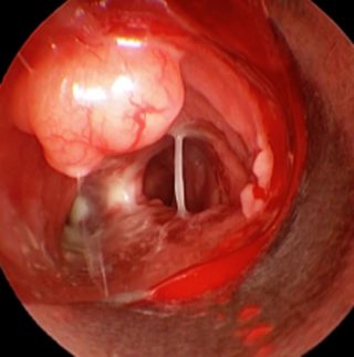
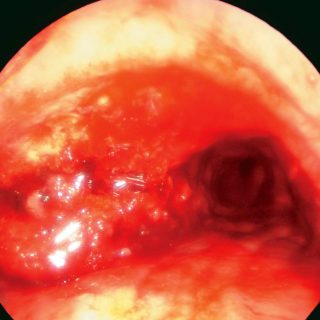
284 – Histoplasmosis

284 - Pulmonary histoplasmo

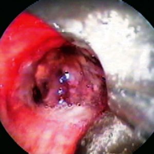
Pulmonary histoplasmosis with bronchial involvement: a voluminous formation occupies the right source bronchus. The carinal edge can be seen at hour 9 of the image. The lesion presents with irregular surface, very vascularized areas and partially covered by retained secretions. The diagnosis was established based on the isolation of the causative agent from the endoscopic … Read more

283 – Tracheal Amyloidosis

283 - Tracheobronchial Amyloidosis

Exophytic lesion that sits on both carinal slopes. The surface is partially impacted and due to its large size it considerably reduces the light of both source bronchi. Changes of coloration in the mucosa with very congested areas and general aspect that makes it similar to a bronchial carcinoma.

280 – Bronchial metastasis. Clear Cell Carcinoma

280 - Endobronchial metastasis

Clear cell carcinoma. A voluminous mass of tissue occupies the light of the right source bronchus, with some protrusions on the surface. Through a triangular light that allows pulmonary ventilation, you can see the entrance of the upper lobe. A plane of separation with the mucosa of the bronchus around the perimeter of the lesion … Read more

279 – Tracheal Metastasis. Carcinoma of Mama

279 - Endotracheal Metastasis

Bronchoscope stopped next to a smooth surface formation but irregular contour, with abnormally voluminous surface vessels. It is an endotracheal metastasis of breast carcinoma. At the bottom of the image, the edema, the enlarged and pale carina, the left main bronchus with its reduced entrance and clogged secretions complete the set of indicative elements of … Read more

276 – Bronchial Carcinoma

276 - Carcinoma

Combination. In the upper part of the image we can see the lesion that is still submucosally located, in the form of a protrusion that seeks to burst into the light, with its plethoric parallel vessels. In the center of the photograph the lesion is vegetative and thrives inside the bronchial lumen. Carcinoma.

275 – Tracheal Carcinoma

275 - Giant cell carcinoma II

Image corresponding to the previous case, after a recanalization of the right source bronchus. There are diffuse inflammatory changes and blood in the path of the bronchus, which can be followed up to the lower lobe. The folds in the source bronchus, deep and tortuous, are diverted to enter the upper lobe, but it is … Read more