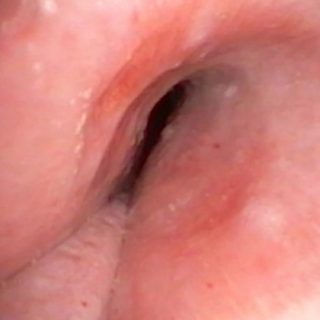
133 – Granuloma
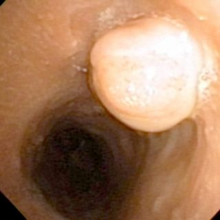
Voluminous and solitary, a pale smooth surface granuloma observes the trachea from its insertion at the edge of the ostoma. Health center Montevideo, Uruguay

Voluminous and solitary, a pale smooth surface granuloma observes the trachea from its insertion at the edge of the ostoma. Health center Montevideo, Uruguay
Lobulated compression on the mucous membrane of the posterior wall of the extrathoracic trachea.
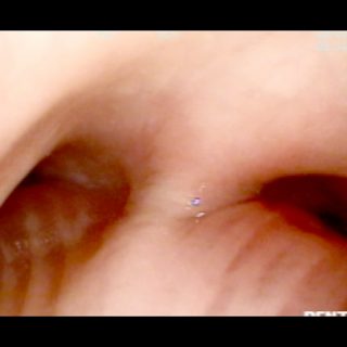
Tracheal carina enlarged and fixed. The entrance to the source bronchi is asymmetric due to unequal reduction in its diameters, due to compression and regular edema.
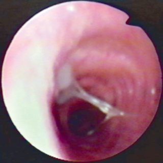
Left lateral compression on the trachea that can only be appreciated by the asymmetry at the source of the bronchial foramen, towards which appears a formation that emerges from the right lateral wall of the trachea and subocludes the entrance to the main bronchus on the same side. Observe the bulging of the posterior mucous … Read more
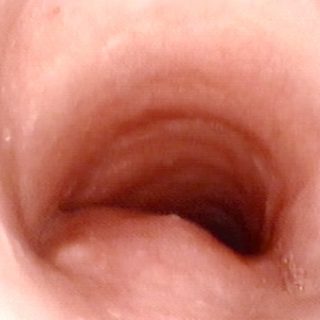
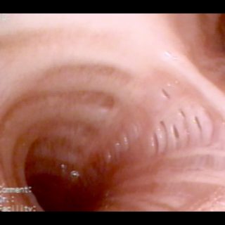
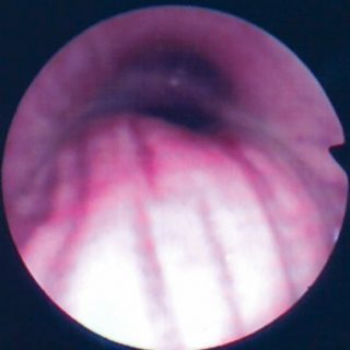
Trachea in “saber scabbard”. Appropriate analogy for this severe and prolonged compression of the trachea by an endothoracic goiter. The cartilaginous reliefs have been lost and the edema gives a regular appearance to the entire mucosa. The rear wall is now a narrow and straight corridor that leads to the carina. Alejandro Posadas Hospital
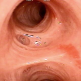
In chronic bronchitis, the atrophy of the mucosa gives it a tense and bright appearance, with wide glandular holes and sharp reliefs on the spurs and cartilages.
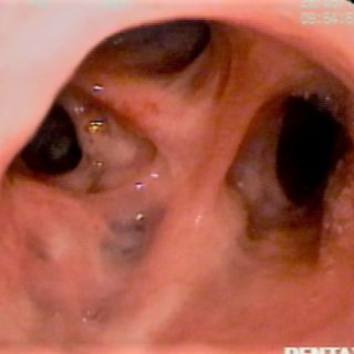
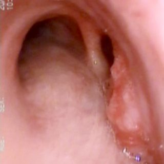
Multiple depressions of the bronchial mucosa in the right upper lobe, with anthracotic macules in them.
Dilated bronchial glands at the entrance of the left source bronchus, at the junction of its lateral wall with the posterior or mucous membrane.
Chronic bronchitis Forward displacement of the posterior wall of the right source bronchus during inspiration. The depth of the longitudinal folds is maintained and does not disappear, as occurs when the wall is “stretched” by extrinsic compression. Here the alteration is dynamic and is due to the flaccidity of the mucosa and the elastic bands.
Chronic bronchitis Thin secretion of mucous aspect in the left source bronchus.