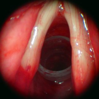
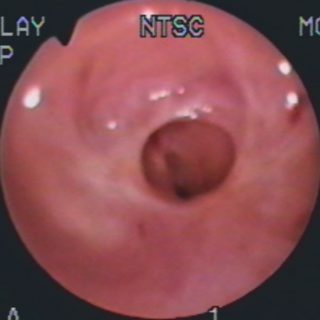
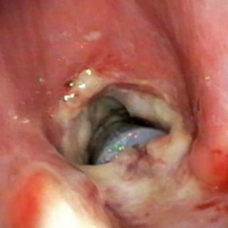
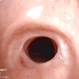
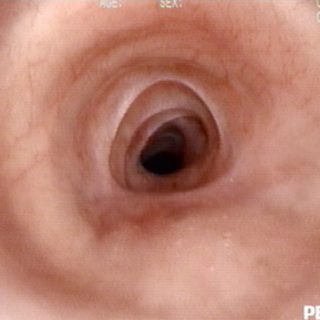
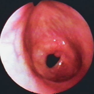
195 – Stent in Tracheal Stenosis

195 - Tracheal stent in unresectable stenosis

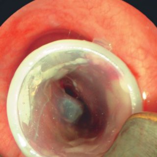
“Ideal” position for a tracheal stent in the treatment of unresectable stenosis. The device is “trapped” in the area of the stenosis and its anterior end is “floating” in the tracheal lumen. Thus, this position reduces the possibility of the appearance of granulomas. The distal end of the prosthesis is still somewhat folded. The defect … Read more

193 – Tracheal Stenosis

193 - Complex tracheal stenosis

Complex tracheal stenosis after endo-surgical treatment. Image taken one month after a stent, that remained for 2 years, was removed. There is sufficient tracheal light and some changes in its relief due to the presence of granulomas on the left wall. The edema erases the longitudinal folds and the silhouette of the cartilages.

190 – Bronchial Stenosis

190 - Benign bronchial stenosis

The light from the left source bronchus is greatly reduced due to a stricture established after a complete bronchial rupture due to chest trauma. The reduction of the bronchial diameter is concentric and progressive or “infundibuliform”. The edema thickens the mucosa and the cartilaginous reliefs and also the linear folds of the posterior wall of … Read more

184 – Subglottic stenosis

184 - Simple stenosis

Subglottis: at short distance from the vocal cords a simple stenosis, in diaphragm, considerably reduces the light. Ventilation is possible thanks to a central and circular hole, with a small marginal nodule. The same volume of inhaled air must increase its speed to enter through a narrow light, becoming turbulent, which causes visible hyperemia in … Read more