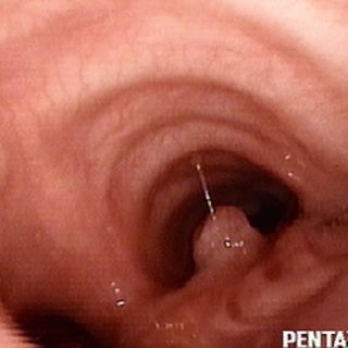
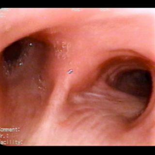
094 – Bronchial Light in “half-moon”
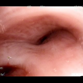
“Light in half moon” Aspect that adopts the bronchial light due to uneven extrinsic compression.

“Light in half moon” Aspect that adopts the bronchial light due to uneven extrinsic compression.
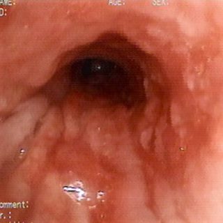
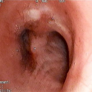
Irregular roughness in the walls of the trachea dominated by the bulging edema, which is missing in the mucous membrane that forms the posterior wall, with its deep folds and loss of its usual parallelism.
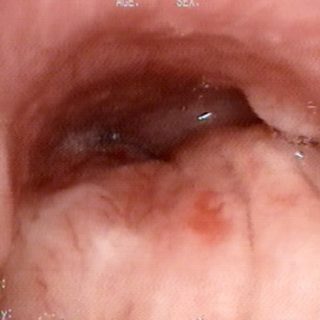
Carina notoriously pathological. Very wide and surely “fixed”. Distortion of the folds that are exaggeratedly marked. On the left, a small submucous protrusion appears at the entrance of the bronchus source.
Locoregional neoplastic disease. Although there is no lesion of endoluminal growth, the bronchial mucosa is decidedly affected by irregular edema and visible thickening. The rigidity and decreased mobility are other signs that can be seen during endoscopy when the bronchus is “fixed” to the tumor that surrounds it.
As a solitary boulder, this carcinoma sits in the center of the right source bronchus, interrupting the normal course of the longitudinal folds.
Full of semiology, the neoplastic repercussion in the interior of the trachea shows deformation of the light at the entrance to the main bronchi, very marked in the left. The carina is widened, with a submucosal protrusion. The folds have deviated and the superficial coloration combines pale areas with congestive ones, in which the full … Read more
View of the entrance to the left source bronchus. In addition to the small protrusion in its right lateral wall, a fine puntilled of submiliar size is observed in the mucosa of the carinal slope.
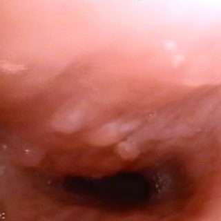
Bronchial light is greatly reduced and deformed by irregular edema of the mucosa in which several mamelons appear on its surface.
Tracheal carina enlarged at the expense of both paths. The biopsy can reveal neoplastic embolisms of the submucosal lymphatics in 11% of the cases.
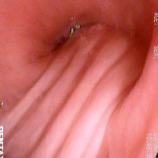
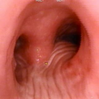
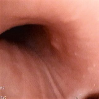
The longitudinal folds are preserved parallel but very accentuated and somewhat “separated” in this pulmonary atelectasis by extrinsic compression. Recall that in the trachea only the malignant conditions contiguous to its posterior wall “accentuate and separate” their folds, while the compressions of nonmalignant causes produce the bulge of the wall and the folds can be … Read more