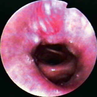
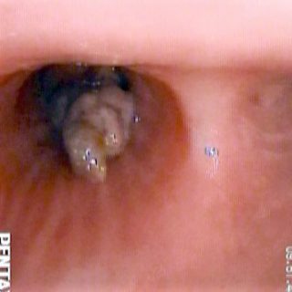
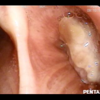
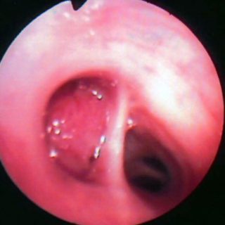
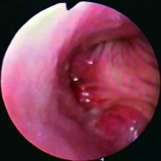
264 – Bronchial Tumor

264 - Sack bottom

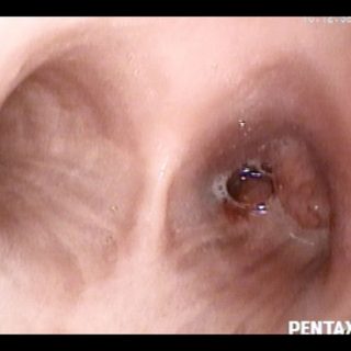
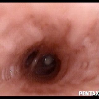
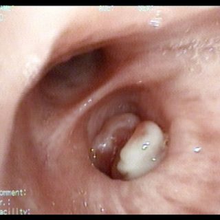
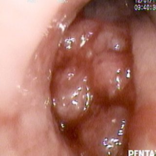
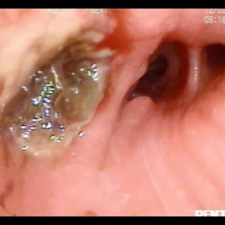
Mucous secretions located in the right source bronchus, which is occluded by a formation that gives it the appearance of “bottom of sac.” This term should be reserved for the description of the bronchial stumps by surgical amputation during pneumonectomies.

263 – Bronchial Tumor

263 - Intermediate bronchus tumor

Although the flexible bronchoscope still runs through the intermediate bronchus, a whitish image due to the small tumor occluding a segment of the base can be guessed in the distance.

262 – Bronchial Carcinoma

262 - Carcinoma

In the form of a double tumor, this vegetative formation of large volume and with one of its ends partially necrotic, arises from the right source bronchus, but is also implanted in the tracheobronchial junction. The biopsy does not present any inconvenience but, naturally, it is necessary to avoid capturing the whitish covering of the … Read more

260 – Bronchial Carcinoma

260 - Small cell carcinoma

Although similar to the previous case, here are indirect signs of neoplastic process, contiguous to the lesion. Irregular edema with some small mamelons on a thickened carinal edge and distortion of the bronchial entrance right source, towards where the longitudinal folds of the posterior wall bend excessively.

258 – Bronchial Tumor

258 - Aged blood clot

This pyramidal formation that emerges from the left source bronchus can be confused with a bronchial tumor, although it is an aged and retained blood clot that acquires this coloration composed of gray and burgundy. Note the normal environment of the surrounding mucosa.

257 – Bronchial Tumor

257 - Necrotic bronchial tumor

A fairly common presentation of large bronchial tumors, in which the surface suffers a necrosis due to being away from its base of implantation, better vascularized, and acquires a whitish creamy appearance.

255 – Bronchial Carcinoma

255 - Undifferentiated carcinoma

View from the left source bronchus, near its fork in the carrefour. A prominent mass rises on its side and bottom walls. The longitudinal folds, very accentuated, lose their parallelism and tend to meet as they enter the upper and lower lobe bronchi. The widening of the secondary carina and the intense edema contribute to … Read more