183 – Blind Trachea

183 - Laryngeal stenosis

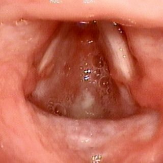
Laryngeal stenosis that has evolved towards the total closure of light at the level of the subglottis. The patient suffers a post-intubation rash with damage to the laryngeal cartilages. It develops a subglottic stenosis that, when it becomes symptomatic or critical, receives the relief of the tracheostomy at the hands of the surgeon. However, this … Read more

180 – Trachea as a bag bottom

a) View of the glottis in abduction. The tracheal lumen is missing at the bottom of the image due to a stenosis. b) Same case. c) Same case: view of the laryngotracheal junction where the complete closure of the light or “blind trachea” can be seen at the level of the first ring.

179 – The middle lobe syndrome

179 - Brock syndrome or middle lobe syndrome

Chest x-ray with loss of volume of the middle lobe in patient with sequelae of tuberculosis and a bronchoscopy that finds the stenosis of the bronchus (upper left quadrant of the image). Brock syndrome or middle lobe syndrome.

178 – Triangular Trachea

178 - Malacias

Triangular light at the site of the old tracheostomy, where the damage or ablation of the cartilage causes a local malacia with total collapse during cough.

177 – Blood Coagulation

177 - Compact clot

This fairly compact clot of incredible size was lodged in the right source bronchus, with the consequent pulmonary atelectasis. In spite of its volume, it was expelled by the pressures generated by the cough.

176 – Endoscopic haemostasis

176 - Endoscopic hemostasis

This small “bronchial plug” of rolled gauze allows to stop a serious hemorrhage when the bronchus is occluded with it. It is applied directly with the rigid bronchoscopy clamp. The long linen thread that holds it will come out through the patient’s mouth and at the moment of removing it, it is only necessary to … Read more

174 – Stony Clot

174 - Blood clot "petrified"

Although the title exaggerates its condition, large blood clots that are not removed in time suffer from drying and hardening that require a rigid bronchoscopy for removal. Most commonly occurs in patients who are under respiratory assistance with insufficiently humidified gaseous mixture.

173 – Hematic Route

173 - Blood in the airway

Line of blood that discovers the bronchus that causes hemoptysis. Sometimes the bronchoscopy can establish the anatomical origin of the hemoptysis, although a trace as clear as the one of the photograph should not be expected. Usually the fresh blood is distributed by the cough and the oldest one coagulates and fragments, so it is … Read more

171 – Blood clot

After a few days, the clot begins to fade, gradually abandoning its original burgundy color. A few more days: before being spontaneously eliminated, the clot, now pale yellowish, retracts, losing volume and increasing its consistency, so that it can be confused with a neoformation.