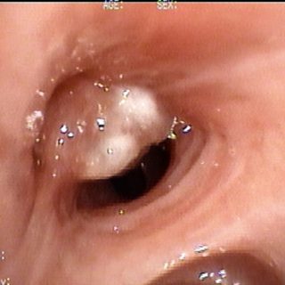
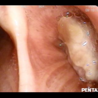
289 – Chondrosarcoma Trachea

As a solitary hump this tumor emerges from the tracheal wall as if it were a uvula. Chondrosarcoma confirmed (Francisco Muñiz Hospital).

As a solitary hump this tumor emerges from the tracheal wall as if it were a uvula. Chondrosarcoma confirmed (Francisco Muñiz Hospital).
This bright, congested and pale tumor, obliterates the entrance of the bronchus culminate in the left upper lobe, allowing only the passage of air to the lingula.
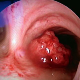
A vegetative formation sits on the right side of the carina. It presents multiple mamelons that give it an irregular and hyperemic aspect that contrasts with the general pallor of the healthy mucosa. An ecchymotic dot is distributed in the area of the edge of the main carina and in its posterior triangle. Squamous cell … Read more
Trachea: a giant cell carcinoma has completely infiltrated the wall. Two ends of the cartilaginous rings are visible in the light as a result of the destruction of their mucosa, now covered by thick purulent secretions and suffering continuous dehydration due to being exposed to the turbulent airflow caused by local narrowness and inflammation.
Complete loss of the normal anatomy of the main carina. View from the distal trachea in which the carina can not be seen, due to the existence of a necrotic surface tissue, with dehydrated purulent secretions that subocludes the entry of both bronchi source: adenocarcinoma.
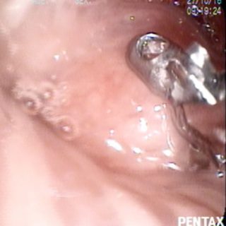
Although the foreground magnifies the image, it is only the small biopsy forceps with fenestrae in its leaflets, commonly used with the flexible bronchoscope, doing its work of tissue capture for the diagnosis of this endobronchial tumor.
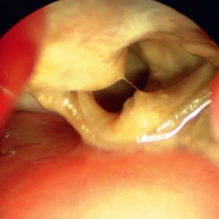
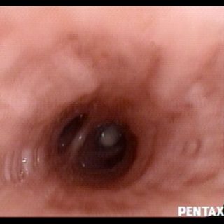
Another vegetative formation that occludes the bronchial lumen but seems “sunken” in the mucosa because it drags the folds towards it.
Tracheal tumor with double origin: two large formations occupy the center of the tracheal lumen, although there is still a sufficient area for ventilation. In the image, the formations appear superimposed, but they have an independent origin constituted by pedicles that emerge from the tracheal wall. Both correspond to a single primary carcinoma.
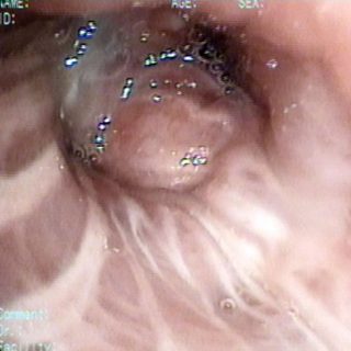
Although the flexible bronchoscope still runs through the intermediate bronchus, a whitish image due to the small tumor occluding a segment of the base can be guessed in the distance.
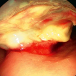
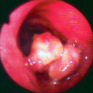
A fairly common presentation of large bronchial tumors, in which the surface suffers a necrosis due to being away from its base of implantation, better vascularized, and acquires a whitish creamy appearance.