190 – Bronchial Stenosis

190 - Benign bronchial stenosis

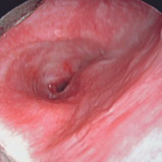
The light from the left source bronchus is greatly reduced due to a stricture established after a complete bronchial rupture due to chest trauma. The reduction of the bronchial diameter is concentric and progressive or “infundibuliform”. The edema thickens the mucosa and the cartilaginous reliefs and also the linear folds of the posterior wall of … Read more

177 – Blood Coagulation

177 - Compact clot

This fairly compact clot of incredible size was lodged in the right source bronchus, with the consequent pulmonary atelectasis. In spite of its volume, it was expelled by the pressures generated by the cough.

112 – Secretions Withheld

112 - passive atelectasis

Mucopurulent secretions. In patients prostrated or with bronchial immobility due to the passive atelectasis that accompanies the effusions, the secretions can fill the lower bronchi, flooding them. You can see the secretions “retained” within the adynamic bronchi that accumulate there due to their decline.

063 – Tracheal Bronchus

063 - Distribution anomaly: tracheal bronchus

Distribution anomaly: tracheal bronchus. An additional bronchus is born in the right side wall and heads to the homolateral upper lobe. Depending on its location, the tracheal bronchus can cause exclusive atelectasis of the superior lobe during anesthetic intubation.