257 – Bronchial Tumor
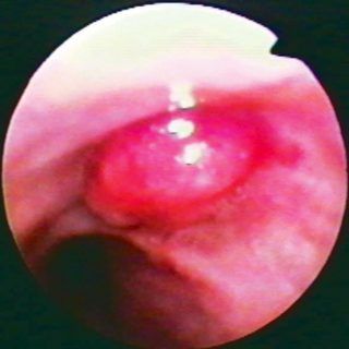
A fairly common presentation of large bronchial tumors, in which the surface suffers a necrosis due to being away from its base of implantation, better vascularized, and acquires a whitish creamy appearance.

A fairly common presentation of large bronchial tumors, in which the surface suffers a necrosis due to being away from its base of implantation, better vascularized, and acquires a whitish creamy appearance.
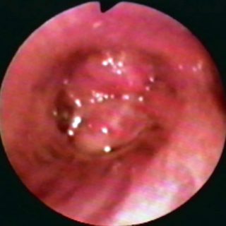
The left upper lobe is occluded by an anomalous tissue, with a homogeneous and somewhat rough surface. The secondary carina looks curiously linear and straight. In the distance you can see the lower lobular, poorly lit.
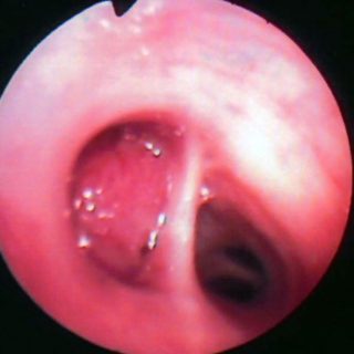
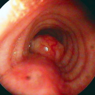
View from the left source bronchus, near its fork in the carrefour. A prominent mass rises on its side and bottom walls. The longitudinal folds, very accentuated, lose their parallelism and tend to meet as they enter the upper and lower lobe bronchi. The widening of the secondary carina and the intense edema contribute to … Read more
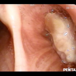
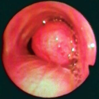
Tumor with “glove finger” appearance. This lesion emerges from the right source bronchus and its independence from the bronchial wall is quite evident. It may have a base of implantation much farther from its visible proximal end. The tracheal carina is pushed to the left.
With a rather classic appearance, this large formation with large submucosal vessels, arises from the posterior tracheal wall and also from the angle that it forms with the lateral walls, as usually happens in adenoid cystic tumors.
The entrance to the upper right lobe bronchus is occluded. In this case, the adenoma presents a familiar aspect. The surface is smooth and a dense submucosal vascular network gives it an intense coloration.
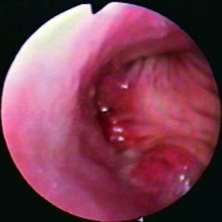
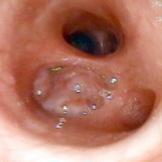
Unlike the previous case, this adenoma that obstructs the left source bronchus presents several protrusions in its surface. Thus, it is easily confused with other types of tumors, especially due to the enlarged appearance of the tracheal carina.
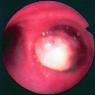
The carial edge can be seen at 9 o’clock. At close range, a large mass occupies the right source bronchus, surmounted by bubbles formed by the instilled saline solution. The formation is smooth, as happens in most adenomas, and the entire mucosa of the surrounding tissue is completely normal.
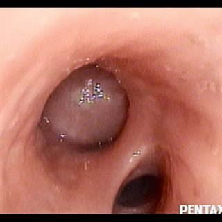
Carcinoid tumor that appears with a fairly classic appearance. Due to its smooth convexity and its free contact with the bronchial walls it is compared with a “glove finger”.
This lesion, somewhat lobed on its surface, completely seals the entrance to the lower lobe bronchus. Photograph of the excised carcinoma.