084 – Bronchial Obstruction
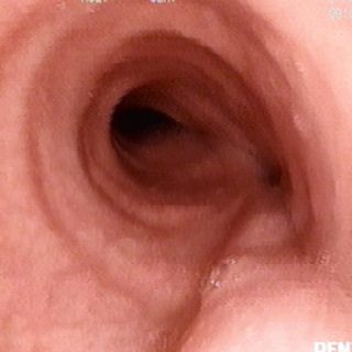
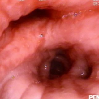
Asymmetry, stenosis and deviation of the marked folds, are the findings in this obstruction of the right source bronchus due to a contiguous extrinsic affection.

Asymmetry, stenosis and deviation of the marked folds, are the findings in this obstruction of the right source bronchus due to a contiguous extrinsic affection.
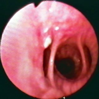
The irregularity of the mucosa, with a granular surface characterizes this neoplastic development in the wall, almost in the center of the image and close to the entrance of the basal segmental bronchi.
Threadlike secretion that crosses the bronchial lumen as if it indicated the area of irregular and mamelonated edema of the mucosa at hour 7: infiltrating carcinoma of the bronchial wall.
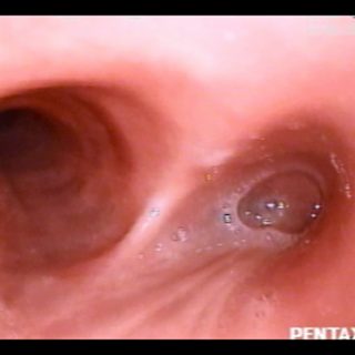
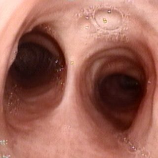
Carina with a rounded edge and widened sheds that give it a “saddle” look. The deformation occurs after the regional ganglion enlargement of different etiologies.
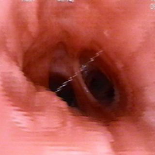
“Hidden carina.” Extensive compression in the tracheal route that produces the prolapse of the posterior wall. Towards the bifurcation deforms the light from the bronchi, losing the right bronchus. The carina it is hidden due to the compression.
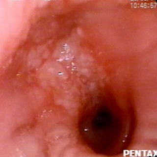
“Stoned edema”. Typical aspect in the left area of the image, where the mucosa, although retaining its homogeneous coloration, is presented as an irregular street on its surface and reminds the stoned of the pavements. It leads here to the anterior segmental of the right upper lobe (RB3) and must be differentiated from the “regular” … Read more
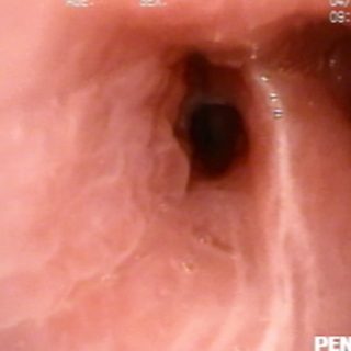
View from the intermediate bronchus. Distortion of the folds at the entrance to the lower lobe, which can be seen at hour 6 of the image. Submucosal miliary infiltration, close to the paracardiac segmental. Habitual aspect in “slit” of its entrance hole.
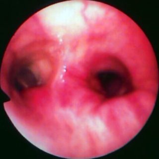
Widening of the left secondary carina: widened and rigid division spur. Local congestion with a sinuous and engorged vessel in the upper part of the image. Distortion of the folds that reach the lower lobe and deformation of its entrance. On the left, the orifice of the lingual bronchus can be seen and the culminar … Read more
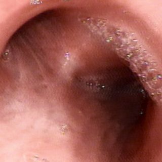
Intense congestive edema of the mucosa that widens the dividing spurs and reduces the light of the segmentals. Also, the longitudinal folds have deepened. Although not exclusive, the finding is common in acute and subacute bronchial inflammatory conditions and the contiguous lung parenchyma.
“Bronchial parallelism”. More common in elders than in the rest of the population, in the parallelism the source bronchi “look straight” to the bronchoscopist.